An MCL injury is a sprain or tear of the medial collateral ligament, which is located on the inner side of the knee. It’s made up of a strong band of connective tissues that connects your femur to your tibia. The MCL works with the other knee ligaments—anterior cruciate ligament (ACL), posterior cruciate ligament (PCL), and lateral collateral ligament (LCL) to stabilize your knee and prevent it from caving inward excessively, like knee valgus.
Causes
MCL injuries are mainly caused by a direct hit from the lateral side of the knee while the foot is planted on the ground. This causes the knee to “cave in” and is quite common in rugby, American and Australian football, and soccer.
Non-contact MCL injuries (valgus stress) are common in skiing and cutting and turning sports like basketball.
ACL vs MCL injury symptoms
Both ACL and MCL injuries have similar symptoms, such as swelling and pain around and inside of the knee. However, an ACL tear has a “popping sound” at the time of the injury while an MCL injury does not.
Other symptoms of an MCL injury include:
- Stiffness and decreased range of motion in the knee
- Instability or feeling like the knee may “give away”
- Locking, catching, or popping sensation in the knee during movement
- Bruising around the inner knee area
Massage therapy for an MCL injury
Massage therapy may alleviate some symptoms of an MCL injury, but that depends on the injury grade. The grades are classified into three levels of severity:
- Grade 1 (Mild): A minor tear where the MCL is overstretched but not significantly damaged. Symptoms include mild tenderness and swelling with little to no instability in the knee.
- Grade 2 (Moderate): A partial tear of the MCL, resulting in more significant pain, swelling, and bruising. The knee may feel somewhat unstable, and there’s usually a noticeable reduction in range of motion and strength.
- Grade 3 (Severe): A complete tear of the MCL, leading to severe pain, swelling, and bruising. The knee is often unstable, and there’s a substantial loss of function and mobility. This grade often requires surgery for a full recovery.
Massage is not recommended for early onset of an MCL injury since it may increase swelling, inflammation, and pain. While there’s no established research to guide and inform therapists and patients how long you should wait, the best approach is to consult with your physician and physical therapist when you may get a massage.
Given the nature of ligament injuries and knee pain, massage therapists could work around the injured area, such as the quadriceps, hamstrings, and calves to alleviate stiffness and decrease pain. Gentle massage techniques should be used, such as Swedish massage and lymphatic drainage massage. A bolster or pillow underneath the knee can help alleviate some tension.
Medial collateral ligament anatomy
Specifically, the MCL is made up of three parts to minimize the risk of getting a MCL injury: the superficial MCL, the deep MCL, and the posterior oblique ligament (POL). These parts are not compartmentalized; rather, they blend together so that the boundaries of each component are not easily noticed.
The superficial MCL attaches at the medial epicondyle of the femur with the semimembranosus tendon, while at the other end of the ligament, it attaches to the medial side and back of the tibia.
The deep MCL is made up of the meniscofemoral and meniscotibial ligaments. The meniscofemoral ligament inserts just below the superficial MCL at the femur and at the medial meniscus. The meniscotibial ligament continues from the medial meniscus to the edge of the medial tibial plateau at the articular cartilage. Although the latter ligament is attached to the meniscus, it offers no stability to the cartilage.
The fibers that line up along the superficial MCL fan out toward the back as it approaches the POL, which also blends into the semimembranosus tendon, medial meniscus, the joint capsule and just below the articular surface of the knee. This section of the knee is called the posteromedial corner. Together, these three parts of the MCL resist forces that push the knee into an excessive valgus position (moving toward the midline of your body) or twist the knee that can cause a MCL injury.
Each layer of the MCL provides a different support in relation to how the knee is flexed or extended. The superficial MCL helps to stabilize the knee when it is externally rotated with the knee flexed at 30 degrees. Meanwhile, the deep MCL helps to stabilize the knee when it is internally rotated throughout the entire range of motion of the knee.
When you flex your knee, the semimembranosus tendon supports the superficial MCL and POL, while the vastus medialis muscle of the quadriceps support the superficial MCL when you extend your knee. How much these supports can help minimize your risk of sustaining a MCL injury would depend on how well-trained you are.
A team of researchers, led by Dr. Milford Marchant, Jr., from Duke University, published a review that mentioned that there is a “wide variation” of how the ligament fibers reinforce the knee joint. This may explain some people with “good muscular support” can continue to perform after an injury and “well-trained athletes” can still “endure higher stressors” at the knee without getting an MCL injury.
MCL injury cannot be fully understood without considering the posteromedial corner since they share the anatomy and movement of the superficial MCL. Marchant, Jr. et al. speculated that the posteromedial corner injuries are studied less because it is usually grouped with MCL injuries. Parts of the posteromedial corner include the POL, semimembranosus tendon, oblique popliteal ligament, and posterior horn of the medial meniscus.
Like the MCL, the posteromedial corner stabilizes the knee when it extends, a common function when you walk or run. The corner slackens when you flex your knee (and the superficial MCL takes over the stabilizing job) or when your tibia externally rotates. In full knee extension, the POL prevents the tibia from sliding back and valgus knee.
The medial articular nerve, which is part of the saphenous nerve that is located toward the end of the femoral nerve, innervates the MCL, while tiny branches of blood vessels and lymph vessels from the superior and inferior genicular arteries provide nutrients and remove metabolic wastes from the ligament.
Sometimes MCL injuries occur with an ACL tear or PCL tear. In cases like these, clinicians must decide whether a patient would need surgery or not as well as risk assessment based on each individual case.
MCL injury diagnosis
In a MCL injury diagnosis, your physician or physical therapist may check your knee’s range of motion, tenderness, pain, and swelling. They may ask you some questions about how the injury occurred and the symptoms you’re experiencing.
A valgus stress test may be used to test for a MCL injury, which tests laxity of the MCL. This is compared with the opposite, uninjured knee. They also may apply gentle stress to your knee joint to see if there is increased laxity or looseness, which could indicate an MCL tear.
While you are lying on the examination table on your back with your injured knee slightly flexed, the clinician would put one hand on your outer knee and the opposite hand on your ankle. Then your knee is gently rotated in and out. Any pain felt may also be a sign to a MCL injury as well as an increase of the joint space at the inner knee.
A positive valgus stress test would be laxity at 30 degrees of flexion, which indicates injury to the superficial MCL. Laxity at 0 degrees of flexion means that the deep MCL is injured.
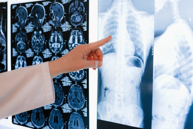
In some cases, they may also use imaging like magnetic resonance imaging (MRI) and an X-ray to check your knee.
Treatment
Initial management of an MCL tear should include controlling pain and swelling and restoring range of motion.
RICE method
Treatment depends on severity of the injury, which may be diagnosed by physical examination but is confirmed with MRI and whether the injury is isolated to the LCL or involves multiple ligaments. All grades of an LCL injury can be treated with:
- RICE method (rest, ice, compression, elevation), especially in the acute phase
- Pain medications (NSAIDs)
- Knee bracing
- Physical therapy, includes exercise
Related: Ice for Pain
Exercise
Exercise may reduce swelling, restores flexibility, strengthens knee muscles for better stability, and prepares the knee for normal activities and sports. Generally, start with low-impact exercises and gradually progress to strengthening and sports-specific activities.
Sample exercise progression may look like this:
Early Phase:
- Quadriceps setting/isometric contraction
- Active straight leg raises
- Heel slides with towel assist (to regain knee flexion)
- Prone hang (to regain knee extension)
Middle Phase (strength and balance):
- Terminal knee extensions with resistance band
- Lateral lunges to single leg balance
- Calf raises
- Leg curls
- Hip abduction
- Hip extension
- Half squats
- Wall slides (mini squats)
Late Phase (higher intensity and range of motion):
- Single leg strengthening progressions
- Leg press
- Hamstring curls
- Knee extensions
- Roman chair (back extensions)
A 2016 MCL injury review, the researchers said that for a grade III injury, movement therapy, quadriceps activation, and regaining a normal gait should be done as soon as possible after a MCL reconstruction surgery. After being in a non-weight bearing hinge brace for six weeks, they suggested that the next steps should be “functional exercise progression” and “proprioceptive-based exercises.” Avoid valgus knee movements during exercise therapy.
Analgesics
Scialoia and Swartzendruber stated that analgesics should be used to manage pain since pain “limits one’s ability to efficiently move the injured area through a full range of motion.” They warned that these painkillers, commonly known as NSAIDS (nonsteroidal anti-inflammatory drugs), block a compound called prostaglandins, which is essential for forming blood clots, controlling infection, and processing inflammation. Thus, long-term reliance on NSAIDS may delay the healing process.
Disclaimer: Follow your physician’s recommendations if you are taking NSAIDS for your MCL injury recovery.
Surgery
While conservative treatments are generally recommended for most people with grade I or II MCL injuries, some research suggests that reconstructive MCL surgery should be used to repair the ligament since non-operative treatments may increase the risk for knee osteoarthritis, valgus knees, and less stability in the knee during rotations.
The type of surgery you may need would depend on the severity of the tear,, the presence of an ACL and/or PCL tear, and other factors that may influence the pain and functional outcomes.
Researchers from the Hospital Universitario La Paz in Madrid, Spain, described several types of MCL surgeries that a surgeon might use.
Primary repair of bone avulsion
Bone avulsion involves using sutures to reattach a ligament to the bone if there’s a bone avulsion—a tear of a piece of bone with the ligament—involved at the tibial or femoral insertion. A surgeon might use anchors and/or staples with the sutures.
Primary repair for MCL entrapment
In a MCL tear, the MCL can be “caught” under the medial meniscus or at the pes anserinus tendons. A surgery would get it out of entrapment and reattached.
Reconstruction
When conservative treatments fail to allow the MCL to heal properly, reconstructive surgery is recommended, which involves using tissues from another part of your body that are similar to the ligament. Sometimes biomaterials (plastic implants that mimic the properties and functions of living tissues) are used instead if grafts from other tissues are not available or if the procedure is too risky.
Please consider that the information about treatment and diagnosis are for educational and informational purposes only, not medical advice.
Further reading
Can a Lateral Meniscus Tear Heal Itself?
How Massage Can Treat Chronic Pain
Massage for Joint Pain: A Biopsychosocial Approach
Nick Ng, BA
Nick Ng is the editor of Massage & Fitness Jounal and the managing editor for My Neighborhood News Group.
An alumni from San Diego State University with a bachelor’s degree in graphic communications, Nick had also completed his massage therapy training at International Professional School of Bodywork in San Diego in 2014. In 2021, he earned an associate’s degree in journalism at Palomar College.
When he gets a chance, he enjoys weightlifting at the gym, salsa dancing, and exploring new areas in the Puget Sound area in Washington state.