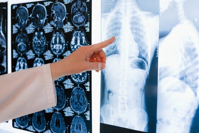
Shoulder impingement is where the tendons and connective tissues in your shoulder get pinched or compressed, causing pain and inflammation. It happens when the space between the bones in your shoulder joint narrows, usually due to repetitive overhead motions or age-related changes. This pinching can irritate the rotator cuff tendons and the bursa (a fluid-filled sac) in your shoulder, leading to pain, especially when you raise your arm overhead or lie on that side. Shoulder impingement is often seen in athletes and people who do a lot of physical work, but it can also develop gradually over time without a clear cause. For years, shoulder impingement has been a stand-alone diagnosis when, in reality, it’s a symptom of another problem. The subacromial space does not shrink on its own; something has to cause the narrowing.
Shoulder anatomy
The shoulder is a complex structure made up of bones, tendons, ligaments, muscles, cartilage, and nerves, with the glenohumeral joint being a key player in its function. Unlike some other joints, like the hip, it doesn’t have a lot of inherent stability, relying instead on strong ligaments and muscles to hold everything together. Think of it like a golf ball balanced on a tee, with the ball representing the arm bone and the tee being the shallow socket of the scapula. The glenoid labrum, a ring of cartilage, adds some depth to this socket, providing a bit more stability. Plus, there are 17 muscles that connect to the scapula, including the rotator cuffs. There’s an extra layer of movement protection afforded by the “roof” of the joint created by the acromion, coracoacromial ligament, and the coracoid process. The space between the roof and the humeral head is the subacromial space, which houses the subacromial bursa and rotator cuff tendons. The insertion of the long head of the biceps tendon is close to this space, but it’s not technically in it. In shoulder impingement, raising the arm compresses the structures in the space between the humeral head and the acromion. Shoulder elevation is created by the interaction of the rotator cuff and stabilizing shoulder muscles as they work to keep the bony anatomy from hitting or rubbing. The rotator cuffs are well-known for their role in throwing, but arguably, their more important job is to maintain the position of the humeral head on the glenoid as it moves beneath the acromion.
How scapulohumeral rhythm affects shoulder impingement
Although there can be some variation in the ratio, it’s widely accepted that there’s about a 2-to-1 relationship between the humerus and scapula during elevation. This is called a scapulohumeral rhythm. Rather than focusing on the numbers, recognize that the scapula is “quiet” during the start of elevation, moves in coordination with the humerus through the mid-range of motion, and then becomes somewhat stable again at angles above 90 degrees. On the back side, the scapular stabilizers control the movement. The upper trapezius, lower trapezius, and serratus anterior are responsible for upward rotation, while the rhomboids, levator scapulae, and pectoralis minor control downward rotation. When your shoulder blade is positioned correctly, it creates more space between the bones of your shoulder joint, known as the acromiohumeral distance. This measurement helps evaluate how much room there is around your shoulder. The occupation ratio percentage takes into account the thickness of certain tissues in the area. A greater acromiohumeral distance and a lower occupation ratio mean there’s more freedom of movement in your shoulder and a lower chance of experiencing impingement issues. Illustration of shoulder impingement.
Types of shoulder impingement
Traditionally, subacromial impingement has been used to describe mechanical encroachment of the structures that live in the subacromial space. Mechanical encroachment is where normal shoulder anatomy “invades” the subacromial space. This occurs during the mid-range of motion and may create a “painful arc.”
External impingement
Subacromial or external impingement is the most common type, also known as swimmer’s shoulder because of the constant repetition swimming entails. This condition is further divided into primary and secondary categories.
- Primary external impingement is where structural changes decrease the subacromial space. Osteophyte formation, variants in acromion type, or an increase in the size of soft tissues can all narrow the space.
- Mechanical impingement is the compression or shear forces to soft tissues. In these cases, it is either a bone or ligament that is impinging on another soft tissue and creating pathological changes.
- Secondary external impingement is where muscle imbalances or tissue tightness can change the shoulder’s movement patterns by changing the humeral head’s position. In these cases, the humeral head moves upward, which decreases the space and compresses the soft tissues.
Internal impingement
Internal impingement is where the undersurface of the rotator cuff tendons are trapped between their attachment on the humerus and the posterior edge of the glenoid. This happens when the arm is at 90 degrees of shoulder abduction and external rotation such that the greater tuberosity of the humerus butts up to the scapula.
Shoulder impingement causes
Shoulder impingement can be caused by many factors, including
- quality of the soft tissues
- age
- flexibility
- anatomical variations
- joint mechanics
- posture
The typical patient with subacromial impingement is over 40-years-old and reports pain without any known trauma. These patients may recall a shoulder injury following repetitive movements, such as gardening and practicing tennis swings. A “painful arc” is generally present when the arm is elevated between 70 and 120 degrees, and pain can prevent them from lying on their side. Rotator cuff pathology can be implicated in primary or secondary impingement. Swelling of the rotator cuff tendons may cause narrowing of the subacromial space in primary impingement. The rotator cuff can be implicated in secondary impingement if weakness interrupts the ability of the muscles to depress the humeral head during arm elevation to steer clear of the acromion. Related: How to Fix Rounded Shoulders
Diagnosis of shoulder impingement
Being diagnosed with shoulder impingement doesn’t always provide the clarity patients are looking for. This can be a “catch-all’ diagnosis that fails to implicate a specific structure which is sometimes frustrating for patients. However, their frustration can be mitigated by patient education. Informing patients about the complexities of shoulder anatomy and biomechanics is often useful to understand why their pain may not stem from a single structure. As with all other muscle and joint conditions, the crux of the diagnosis of impingement is a thorough history and physical examination. Patients may present with posterior shoulder stiffness or instability. Special attention should be paid to the scapulohumeral rhythm to identify any abnormal movement. Hypermobility or instability of the glenohumeral joint should also be noted.
Shoulder tests
Four shoulder tests are commonly used to see if impingement is present, but none are particularly good at pinpointing what structure is being impinged.
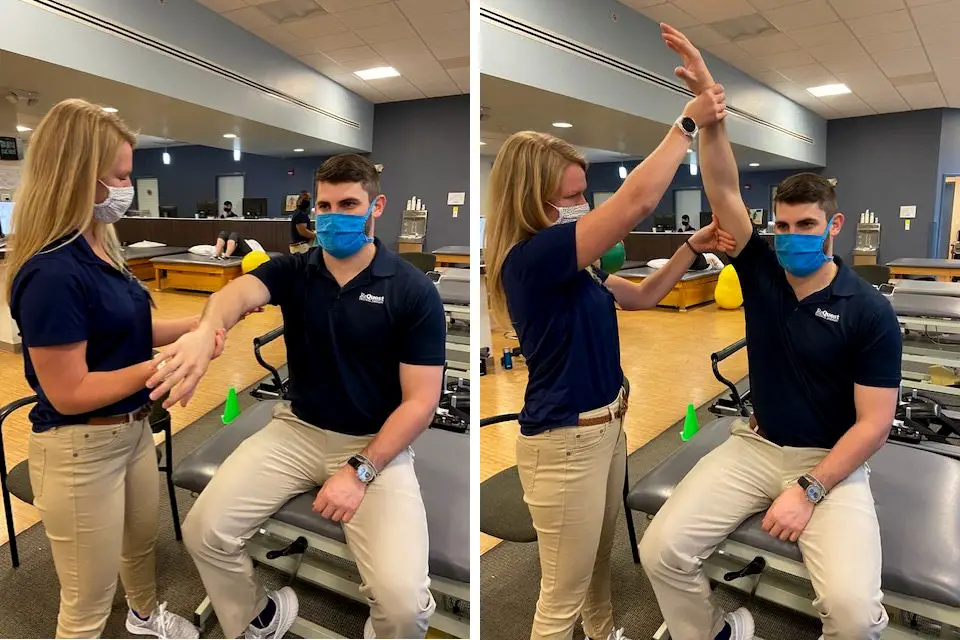
Neer test
The Neer test is performed by stabilizing the scapula with one hand and then passively raising the patient’s arm anteriorly with the other hand. This motion narrows the subacromial space, which can provoke pain if there is impingement of the rotator cuff tendons or other structures The Neer test evaluates for impingement of the supraspinatus tendon against the anterior third of the acromion, coracoacromial ligament, or other structures in the subacromial space. Anterior pain indicates subacromial impingement while posterior pain indicates internal impingement.
Jobe Test The Jobe test is performed by abducting the scapula to 90 degrees in the scapular plane with the elbow flexed at 90 degrees. The patient’s arm is then fully internally rotated, with the thumb pointing down, as if emptying a can. The examiner applies a downward force on the distal arm while the patient resists. A positive Jobe test is indicated by pain, weakness, or the inability to resist the downward force on the affected shoulder compared to the unaffected side. This suggests supraspinatus muscle weakness or irritation, which can be a sign of subacromial impingement.
Hawkins-Kennedy test
The Hawkins-Kennedy test is performed by placing the arm at 90 degrees of forward flexion with the elbow flexed to 90 degrees. The examiner then stabilizes the scapula with one hand and uses the other hand to passively internally rotate the patient’s arm. A positive test is indicated if there’s pain during this internal rotation maneuver. The Hawkins-Kennedy test is thought to narrow the subacromial space and potentially impinge the rotator cuff tendons, particularly the supraspinatus and subscapularis. However, the test has been shown to have relatively high sensitivity (79%) but low specificity (59%), meaning it may not be highly accurate in isolating subacromial impingement. Sensitivity measures how well a test correctly identifies those with a condition, while specificity measures how well it correctly excludes those without the condition.
Scapular assistance test
This scapular assistance test (SAT) test is performed by stabilizing the scapula by fixing the clavicle and scapular spine with one hand, and grabbing the inferior angle of the scapula with the other hand. The patient then performs arm elevation or abduction, and the examiner assists the movement of the scapula. A positive SAT is indicated if the patient experiences less pain during the assisted movement compared to the unassisted movement. It also suggests weakness or poor activation of the scapular stabilizers, such as the serratus anterior and lower trapezius muscles. The SAT can help determine if scapular stabilization exercises should be included in the rehab program for the patient. Although most patients with impingement don’t need imaging, it can provide insight into the problem. Radiographs are used to examine the shoulder, like measuring the acromiohumeral distance. In most adults, this distance is 10 millimeters. A narrowed distance, typically less than 6 to 7 millimeters, is indicative of subacromial impingement, rotator cuff tear, or tendinopathy.
Conditions that mimic shoulder impingement
The differential diagnosis of shoulder impingement is complicated because there are several underlying pathologies that may be related to impingement. Clinicians need to rule out:
- frozen shoulders
- rotator cuff tears/tendinopathy
- scapular dyskinesia
- instability, biceps tendonitis
- labral tears
The signs and symptoms of most shoulder conditions are quite similar. Just to muddy things up a little more, pain may be caused by more than one structure or pathology. Patients with shoulder conditions may report the following regardless of diagnosis:
- Pain with overhead motion
- Pain when reaching out to the side
- Inability to lie or sleep on the involved side
- Tenderness at the front of the shoulder or mid-humeral region
- Aching at night
- Pain when reaching behind the back
- Weakness or stiffness of the involved side
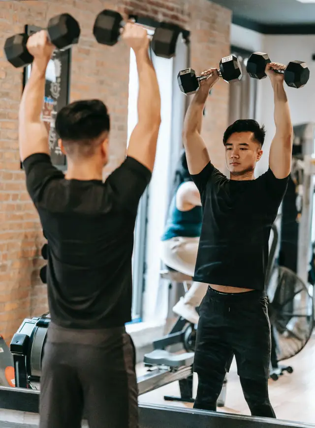
Shoulder impingement treatments
There’s strong scientific evidence and anecdotal support for delaying surgery in favor of conservative management. Despite a lack of research support, subacromial decompression (acromioplasty) surgery remains the most common arthroscopic procedure at the shoulder. A 2020 systematic review found “high certainty evidence of no additional benefit of subacromial decompression surgery over placebo surgery” in reducing pain one year after the surgery. A period of three to six months of conservative management should be employed before you even consider surgery. The patient should be educated on activity modification that includes avoiding overhead movement, heavy mechanical loading, and repetitive motion. A 2017 systematic review and meta-analysis concluded that exercise should be used in the treatment of this condition, and non-steroidal anti-inflammatories and corticosteroids were better than placebo for controlling pain. Although exercise can treat shoulder pathology, the type and amount of exercise have not yet been established.
Exercises
Shoulder impingement exercises typically involve targeting the rotator cuff and scapular stabilizers. However, there is not enough good quality evidence that exercises that target specific muscles (e.g. rotator cuffs) are better than general exercises (e.g. jogging, lat pulldowns, push-ups). And so, exercise recommendations should also factor patients’ preferences and abilities.
Bilateral external rotation
– Sit or stand with a tall posture and your shoulder blades set down and back (like you’re putting them in your back pockets). – Bend your elbows to 90 degrees and pinned to your ribs. – Hold a resistance band, dumbbell, soup can, or similar objects for resistance with your palms facing up. – Pull your hands apart (thumbs go in opposite directions). – Pretend like your hands are full of soup that you don’t want to spill. They should stay parallel to the ground through the full available range of motion and then come back together in the center of your body.
T exercise
– Use a counter, table, or back of the couch—something that can safely support your body weight. – Hinge forward at your hips and support yourself on the forearm of your non-involved shoulder. The exercise starts with your involved side hanging straight down so that your fingers are pointing toward the floor and your palm is facing your midline. – Set your shoulder blade down and back, and then raise your arm up until it is parallel to the floor. Your arm should come straight up so that if both arms were in this position you would look like the letter “T.” A common error in this exercise is to let the bigger, stronger muscles take over and raise your arm so that your hand ends up close to your hip. – Your hand should be in-line with your shoulder in the finish position.
Prone W
– Lie on your stomach with your arms comfortably bent by your sides and your hands in-line with your ears. – With your shoulder blades down and back, raise your arms off the floor until they are even with your body. From a birds-eye view, it should look like you are making the letter “W” with your arms and head.
Reverse kettlebell carry
– Hold a kettlebell or dumbbell over your head with your arm out straight. Your palm should face forward as if you are a waiter carrying a high tray. – Engage your abdominal muscles and hold the weight steady while you walk for about 30 seconds. If you have to arch your back to hold the weight up, it’s too heavy. Researchers in a 2017 study wanted to find out what type of progressive loading exercises were most effective. A panel of experienced therapists chose three loading strategies after an extensive literature review:
- minimally loaded range of motion exercises
- open kinetic chain loading
- closed kinetic chain loading
All three groups had reduced pain and improved function, but no group was better than another. This may suggest that simply doing something to load the rotator cuff is the most important guideline to follow.
Massage for shoulder impingement
It is generally okay to get massage therapy for shoulder impingement, but take precautions before you get one. While massage therapy can be an effective treatment for reducing shoulder pain and improving range of motion, avoid deep tissue massage if the shoulder pain is acute and inflamed. Patients with certain medical conditions like uncontrolled high blood pressure or blood clotting disorders may need to avoid or modify massage. Finally, listen to your body during massage and communicate with the therapist if any movements or pressure cause significant pain. Further reading Shoulder Abnormalities May Not Always Be a Cause of Pain or Disability Descending Modulation: Why Massage Therapy Can Help Alleviate Pain How Massage Therapy Treats Chronic Pain

Penny Goldberg, DPT, ATC
Penny Goldberg, DPT, ATC earned her doctorate in Physical Therapy from the University of Saint Augustine and completed a credentialed sports residency at the University of Florida. She is a Board Certified Clinical Specialist in Sports Physical Therapy.
Penny holds a B.S. in Kinesiology and a M.A. in Physical Education from San Diego State University. She has served as an Athletic Trainer at USD, CSUN, and Butler University.
She has presented on Kinesiophobia and differential diagnosis in complicated cases. Penny has published on returning to sports after ACL reconstruction and fear of movement and re-injury.
Outside of the clinic, Penny enjoys traveling, good cooking with great wine, concerts, working out and playing with her dogs.